


In the case of thoracic osteochondrosis, the organs associated with areas of the spinal cord, located at the level of the affected thoracic region and below, often suffer. Violation of the normal activity of the spine leads to immobility of the arms, legs and torso as a whole, dysfunction of the pelvic organs, respiratory muscles and internal organs.
Osteochondrosis is a degenerative-dystrophic disease of the spine, which is based on a change of the intervertebral discs with involvement in the pathological process of the adjacent vertebrae and intervertebral joints with the entire ligament apparatus.
Characteristics of the anatomy of the spine
The mobility and stability, elasticity and resilience of the spine depend to a large extent on the intervertebral discs, which are one of the types of cartilaginous connections between the bones and provide a strong bond between the bodies of the adjacent vertebrae. The total length of the intervertebral discs is one quarter of the length of the spine.
The most important function of discs is to reduce the vertical load on the vertebrae. The disc consists of three parts:
- vitreous plaques (tightly next to the vertebrae).
- pulposus nucleus (fills the gap between the plates).
- fibrous ring (surrounds the nucleus from the outside).
The nucleus contains cartilage cells, closely intertwined collagen fibers and chondrin (proteoglycans). The anterior surface of the discs is covered by the anterior longitudinal ligament, which is tightly attached to the vertebrae and rotates freely over the discs. The posterior longitudinal ligament fuses firmly with the surface of the disc and forms the anterior wall of the spinal canal. The intervertebral disc does not have its own blood supply, so it feeds on substances that come from diffusion from the vertebral bodies.
The distribution of vertical loads in the spine occurs due to the elastic properties of the discs. As a result of the pressure, the pulp core expands and the pressure is redistributed to the fibrous ring and the vitreous plates. During movement, the nucleus moves in the opposite direction: when it bends - towards the curvature, when it does not bend - forward. When the spine moves, muscles, ligaments and discs are involved in the work. Therefore, a breach in a link leads to a breach of the entire kinetic chain.
Causes and mechanism of development of the disease
In the development of osteochondrosis, a special role is played by the mechanical effect on the spine. Under the influence of adverse static and dynamic loads, the pulp nucleus gradually loses its elastic properties (as a result of depolymerization of polysaccharides), forming protrusions and isolations.
The process of disc degeneration is influenced by a genetic predisposition, which causes changes in the neuromuscular apparatus of the back, a change in the structure of glucosamines and a violation of the distribution of collagen fibers in the disc. The genetic factor is primary in the occurrence of thoracic osteochondrosis, under the condition of increased functional activity.
Risk factors for the development of degenerative changes in the spine include the anatomical features of the discs, which are developmental defects. One of these characteristics is the nutritional characteristics of the structures. In the human body, the disc is made up of tissue with poor diffusion. Closure of blood vessels already occurs in childhood. Post-feeding occurs due to the diffusion of substances through the end plates.
Nutrient penetration stimulator is a dosing load that eliminates static postures and high stress. Physical inactivity is one of the main risk factors for thoracic osteochondrosis. Therefore, regular exercise is an important preventive measure.
The peculiarity of the microscopic structure - a few cells - reduces the intensity of the regenerative capacity and the recovery rate of the disc components. Anatomical feature is the weakness and lack of endurance of the discs in the posterior incisions. This contributes to the appearance of wedge-shaped discs in the lower thoracic and lumbar regions.
Great importance in the development of osteochondrosis is given to the convergent changes. Active degenerative changes begin to increase after 30 years. The synthesis of the ingredients necessary for the disc (glycosaminoglycans) continues, but their quality deteriorates. Hydrophilia decreases, fibrous texture increases, sclerosis occurs.
Stages of degeneration of the intervertebral discs:
- prolonged asymptomatic course, degenerative changes in the endotracheal components, displacement of the nucleus within the disc.
- Severe radical symptoms of thoracic osteochondrosis, compression of the spinal cord, protrusion of the pulpal nucleus (protrusion, 1 degree).
- disc herniation with hernia protrusion (2nd degree hernia).
- degenerative changes in extra-disc components (grade 3).
The pathological protrusion compresses the nerve roots, blood vessels or spinal cord at various levels (cervical, thoracic, lumbar), which determines the clinical picture.
Restriction of mobility in the thoracic spine, which is due to the presence of the thorax, contributes to less injury to the intervertebral discs, and therefore to osteochondrosis. Normal thoracic kyphosis contributes to the redistribution of the weight of the upper half of the body to the lateral and anterior parts of the vertebrae. As a result, intervertebral hernias and osteophytes form on the anterior and lateral surface of the spine. Posterior osteophytes and hernias are extremely rare.
Osteochondrosis contributes to the narrowing of the intervertebral foramina and the compression of the spinal cord roots and sympathetic fibers. The sympathetic fibers originate from the gray matter of the spinal cord, then accumulate in nodes, from which they are sent to all the internal organs. This leads to the fact that thoracic osteochondrosis, in addition to typical neurological disorders, leads to dysfunction of internal organs (stem, vasomotor, trophic) and imitation of physical diseases. This feature of osteochondrosis of the thoracic discs explains the difficulties in diagnosing and prescribing the right treatment.
Symptoms of thoracic osteochondrosis
Chest osteochondrosis is more common in people with a sedentary lifestyle. At the same time, there is no stimulating effect of the dosing loads on the spine, which helps to stop the recovery of the disc. Diseases develop in people who work on a computer for a long time, crouch, etc. such people must perform therapeutic exercises independently.
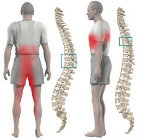
Most often, osteochondrosis of the chest is manifested by dull aches, less often pain and burning. The pain is located between the shoulders. The patient is bothered by the feeling of compression of the chest. When you feel the thorny processes of the thoracic vertebrae, local pain is detected, which increases with axial loads on the spine, deep inhalation and twists of the body.
Some patients have acute pain in the shoulder and lower chest (posterior lateral syndrome). This symptomatology develops as a result of displacement of the lower ribs. The pain increases sharply as the torso rotates. Most often, the pain syndrome disappears abruptly.
Often the chest pain becomes a belt, corresponding to the course of the intercostal nerve. Sensitivity in the nerve zone of the respective nerve ending is disturbed, paraesthesia occurs and a decrease in superficial and deep sensitivity is often observed. Possible violation of the function of the abdominal press, change in the knee and tendon reflexes of the heel.
Violation of the function of internal organs occurs when any nerve root is compressed at the level of 1 to 12 chest. In the thoracic region there are structures responsible for the innervation of the lungs, heart, intestines, liver, pancreas and kidneys. Therefore, there are no characteristic signs only for thoracic osteochondrosis.
The disease is manifested by symptoms that are characteristic of another pathology:
- difficulty breathing?
- intense night pains;
- "heart", angina pectoris.
- pain in the mammary glands.
- pain in the right or left hypochondrium (symptoms of cholecystitis and pancreatitis).
- pain in the throat and esophagus.
- epigastric pain in the abdomen (symptoms of gastritis, enteritis and colitis).
- sexual dysfunction.
Diagnostics
Chest x-ray has the greatest value in the diagnosis of thoracic osteochondrosis. The image shows a decrease in the height of the intervertebral disc, hardening of the end plates, formation of osteophytes.
Computed tomography allows you to determine the condition of the vertebrae, the joints of the spine, the size of the spinal canal, determine the location of the hernia protrusion and its size.
When conducting the differential diagnosis, it is necessary to carefully collect a history and compare all the clinical signs of thoracic osteochondrosis with symptoms of other diseases. For example: heart pain with osteochondrosis does not stop with nitroglycerin, epigastric pain is not related to food intake, it is not seasonal, all symptoms appear mainly at night and disappear completely after a night's rest.
How to treat thoracic osteochondrosis?
Treatment of osteochondrosis of the thoracic spine in almost all cases is conservative. The indication for treatment is the predominance of visceral syndromes with neurological disorders. The main orthopedic treatment should be adequate traction of the spine:
- active vertical traction under water.
- passive horizontal traction on a sloping bed using the Glisson loop in case of damage at the level of 1-4 thoracic vertebrae, from the axillary straps in case of damage at the level of 4-12 thoracic vertebrae.
Medication consists of performing paravertebral blockades with novocaine solution. As the disease worsens, analgesics and sedatives are used. With an unexplained pain syndrome, the use of ointments with analgesic and anti-inflammatory drugs at home is allowed.
After eliminating the acute effects, massage of the muscles of the back and lower extremities is used. Manual therapy is indicated for 1-3 degrees of osteochondrosis in case of development of functional blockages. Includes various options for smooth and rough results on the back muscles.
Therapeutic exercise allows you to load all parts of the spine in a dosing manner, which stimulates the recovery process. An important prerequisite for exercise therapy for osteochondrosis is the exclusion of vertical loads.
Physiotherapy: UHF therapy, ultrasound, inductive heat, radon and pine-conifer salt baths. At the spa stage, active underwater attraction and hot tub are used.
Surgical treatment is rarely used. The indication for surgery is the compression of the spinal cord by prolapse of a disc fracture.